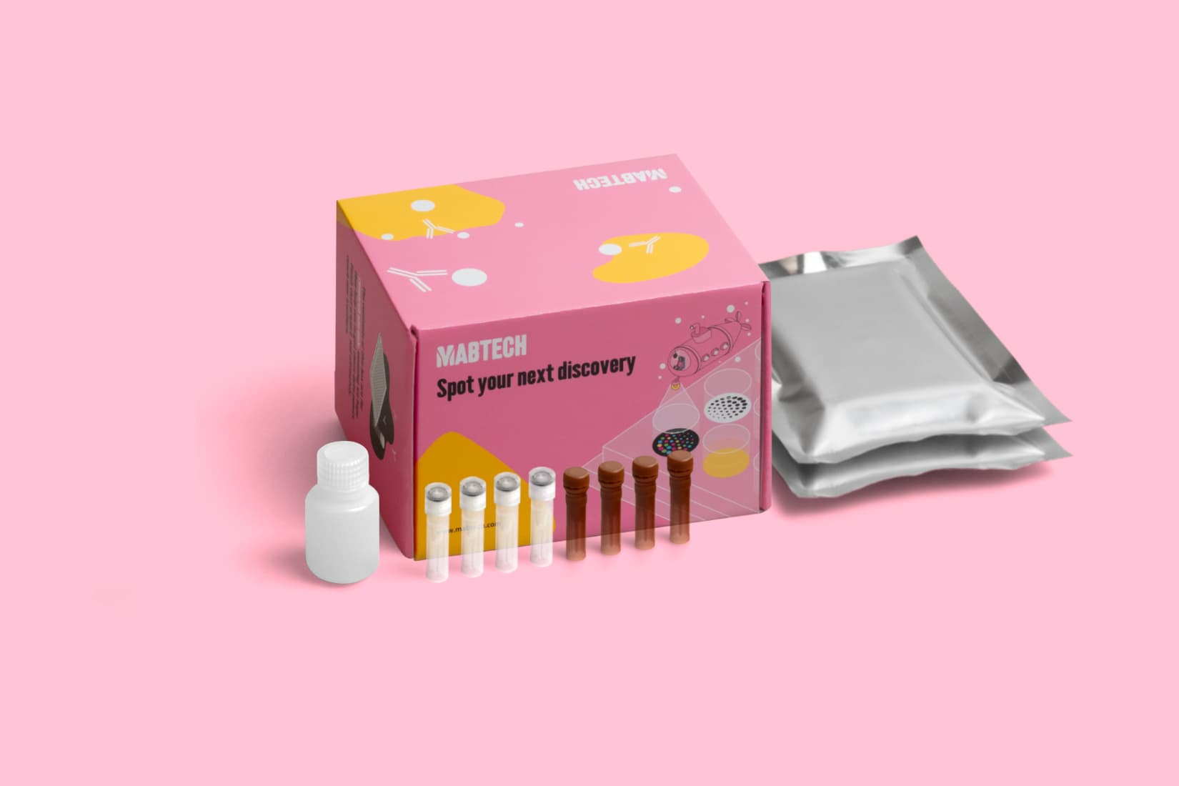
FluoroSpot Plus: Human IL‑6/IL‑1β/GM‑CSF/TNF‑α
FluoroSpot Plus: Human IL‑6/IL‑1β/GM‑CSF/TNF‑α
Special offer
You may add any of these complementary products at a reduced price.
- Reactivity:
- Application:
- Plates:
$300
$240- Reactivity:
- Application:
- Plates:
$245
$196- Reactivity:
- Application:
- Plates:
$545
$436This offer is valid when purchasing FluoroSpot Plus: Human IL-6/IL-1β/GM-CSF/TNF-α or other qualifying products.
Components
| Plate | Pre-coated FluoroSpot plate (mAbs 13A5, MT175, 21C11, and MT25C5) |
| Detection mAbs | Anti-IL-6 mAb (39C3), DIG |
| Anti-IL-1β mAb (7P10), BAM | |
| Anti-GM-CSF mAb (23B6), biotin | |
| Anti-TNF-α mAb (MT20D9), WASP | |
| Fluorophore conjugates | Anti-DIG mAb, 380 |
| Anti-BAM mAb, 490 | |
| SA-550 | |
| Anti-WASP mAb, 640 | |
| Buffer/Solution | FluoroSpot enhancer |
In stock
Delivery 4-9 business days
Shipping $0
Complementary products
Complementary products
IL-6
| Analyte description | Interleukin 6 (IL-6) is a pleiotropic cytokine produced by many different cell types and plays a role in a wide range of functions, such as immune responses, acute-phase reactions, and hematopoiesis. Among other things, it augments antibody production from activated B cells in vitro. |
| Alternative names | Interleukin 6, IL-6, IL6, IFB-B502, BSF-2, BCDF, BSF2, CDF, HGF, HSF, IFN-beta-2, IFNB2 |
| Cell type | B cell, Monocyte/MΦ, mDC |
| Gene ID | 3569 |
IL-1β
| Analyte description | Interleukin 1ß (IL-1ß) is a proinflammatory cytokine and inducer of acute phase responses. IL-1ß is produced primarily by monocytes, macrophages, and dendritic cells after induction by microbes. |
| Alternative names | Interleukin-1ß, IL-1ß, IL-1F2, Interleukin-1beta, IL-1 beta, IL1b, Interleukin-1 beta, IL-1, IL1-BETA, IL1F2, IL1beta |
| Cell type | Monocyte/MΦ, mDC |
| Gene ID | 3553 |
GM-CSF
| Analyte description | Granulocyte macrophage-colony stimulating factor (GM-CSF) can be secreted by T cells, macrophages, endothelial cells, and fibroblasts. GM-CSF stimulates the survival and functional activities of myeloid, that is, monocytes, macrophages, DCs, neutrophils, and eosinophils. It also stimulates the differentiation and proliferation of hematological progenitors. |
| Alternative names | Granulocyte macrophage-colony stimulating factor, GM-CSF, CSF2, CSF, GMCSF |
| Cell type | T cell |
| Gene ID | 1437 |
TNF-α
| Analyte description | Tumor necrosis factor (TNF), also known as TNF-α, is produced by many different cell types, e.g., monocytes, macrophages, T cells, and B cells. Among the many effects of TNF-α are protection against bacterial infection, cell growth modulation, immune system regulation, and involvement in septic shock. |
| Alternative names | Tumor necrosis factor-α, TNF-α, TNF-alpha, TNF-a, TNFa, Tumor necrosis factor-alpha, TNF, DIF-alpha, TNFA, TNFSF2, TNLG1F |
| Cell type | T cell, Tc, Th1, Th2, Th17, Tfh, Monocyte/MΦ |
| Gene ID | 7124 |
