ELISpot Path: SARS‑CoV‑2 (RBD) Human IgG (ALP)
ELISpot Path: SARS‑CoV‑2 (RBD) Human IgG (ALP)
Publications: 26
Documents
Bulk or custom format
A straightforward setup for functional analysis of a specific scientific question.
Components
| Plate | Pre-coated ELISpot plate, white MSIP (mAbs MT91/145) |
| Detection mAb | MT78/145, biotin |
| Antigen | RBD-WASP |
| Enzyme conjugates | Streptavidin-ALP enzyme conjugate for ELISpot |
| Anti-WASP mAb, ALP | |
| Substrate | BCIP/NBT-plus substrate for ELISpot |
| Buffer/Solution | Reconstitution buffer |
In stock
Delivery 4-9 business days
Shipping $0
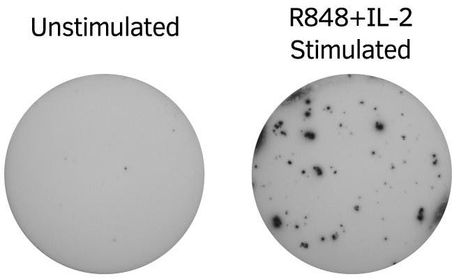
Human PBMC from a COVID-19 convalescent individual were incubated in the presence and absence of R848+IL-2 (stimulates memory B cells) in tubes for three days. Cells were then extensively washed and added to an ELISpot plate (200,000 cells/well) coated with anti-human IgG mAbs. RBD-specific spots were detected with RBD-WASP, i.e., recombinant Receptor Binding Domain protein with a WASP-tag, followed by anti-WASP-ALP and the number of cells secreting RBD-specific IgG was analyzed by ELISpot.


Loading publications...
IgG
| Analyte description | Immunoglobulin G (IgG) is the most abundant Ig isotype in serum, making up approximately 80% of all serum immunoglobulins. In humans, there are four subclasses of immunoglobulin G, with the highest serum concentrations of IgG1 followed by IgG2, IgG3, and IgG4. In mice, the IgG subclasses are defined as IgG1, IgG2a/c, IgG2b, and IgG3. The IgG molecule consists of two heavy and two light chains (κ or λ), resulting in a molecule with two arms for antigen binding. High levels of IgG antibodies are induced following the initial IgM response in a typical immune response to antigens. |
| Alternative names | Immunoglobulin G, IgG, IgG3 |
| Cell type | B cell |
| Gene ID | 3500, 3501, 3502 |
1 / 1