FluoroSpot Path: SARS-CoV-2 (Spike+RBD) Human IgG

FluoroSpot Path: SARS-CoV-2 (Spike+RBD) Human IgG

FSP-05R-RS1-1

Publications: 0
Documents
Questions?
Paves your way to functional analysis of a specific research question.

Components

Detection mAbAnti-IgG mAbs (MT78/145), 550
Fluorophore conjugatesAnti-WASP mAb, 640
Anti-GAL mAb, 490
AntigensSpike-GAL
RBD-WASP
StimuliR848
Recombinant human IL-2
PlatePre-coated FluoroSpot plate (mAbs MT91/145)
Buffers/SolutionsFluoroSpot enhancer
Reconstitution buffer

In stock

Delivery 4-9 business days

Shipping $0

Product specifications

Intended use

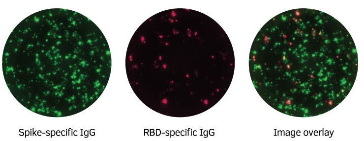
This FluoroSpot Path kit is intended for the enumeration of B cells secreting human IgG in response to SARS-CoV-2 (Spike and RBD) using the FluoroSpot assay. B cells secreting IgG irrespective of antigen specificity (total IgG) can also be enumerated. For research use only. Not for use in diagnostic procedures.

Serum/Plasma samples

For memory B cells, we recommend pre-stimulation with R848 and recombinant human IL-2 (both included).

Recommendation

Product details

ProductFluoroSpot Path: SARS-CoV-2 (Spike+RBD) Human IgG
ApplicationFluoroSpot
AnalyteIgG
ReactivityHuman
Specificity

This kit is based on a matched pair of mAbs specific for the Fc part of human IgG.

Shipping and Storage

Shipping

Shipped at ambient temperature.

Storage

Store antibodies, fluorophore conjugates, and FluoroSpot enhancer at 4-8 °C upon receipt. Recombinant proteins should be stored at -20 °C or below. Plates may be kept at room temperature.

Shelf lifeAt least 12 months from date of receipt.
Human PBMC from a COVID-19 convalescent individual were incubated with R848+IL-2 (stimulates memory B cells) in tubes for three days. Cells were then extensively washed and added to an FluoroSpot plate (250,000 cells/well) coated with anti-human IgG mAbs. Spike-specific IgG spots were detected with Spike-GAL, i.e., recombinant Spike protein with a GAL peptide tag, followed by anti-GAL-490. RBD-specific spots were detected with RBD-WASP, i.e., recombinant Receptor Binding Domain protein with a WASP peptide tag, followed by anti-WASP-640. Spots were analyzed with Mabtech IRIS.

Tutorial, Published November 24, 2022

Cell stimulation in ELISpot and FluoroSpot
In this feature, we’ll walk you through the optimal use of different polyclonal and antigen-specific stimuli.
Loading publications...

IgG

Analyte description

Immunoglobulin G (IgG) is the most abundant Ig isotype in serum, making up approximately 80% of all serum immunoglobulins. In humans, there are four subclasses of immunoglobulin G, with the highest serum concentrations of IgG1 followed by IgG2, IgG3, and IgG4. In mice, the IgG subclasses are defined as IgG1, IgG2a/c, IgG2b, and IgG3. The IgG molecule consists of two heavy and two light chains (κ or λ), resulting in a molecule with two arms for antigen binding. High levels of IgG antibodies are induced following the initial IgM response in a typical immune response to antigens.

Alternative namesImmunoglobulin G, IgG, IgG3
Cell typeB cell
Gene ID3500, 3501, 3502