
FluoroSpot outperforms ICS in a patient-derived lung cancer model
Published: July 27, 2026
7 minute read
Authored by: Jens Gertow
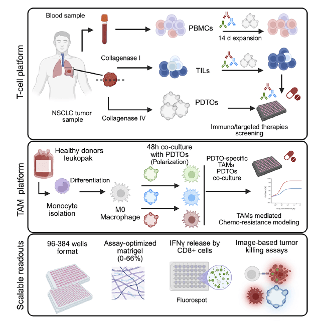
Researchers at Weill Cornell Medicine and Eli Lilly set out to build a patient-derived tumor organoid (PDTO) platform for non-small cell lung cancer (NSCLC) that could model the real tumor immune microenvironment (TIME) and test checkpoint inhibitor combinations on patient material. At the center of their immune-monitoring strategy was our FluoroSpot Plus: Human IFN-γ/Granzyme B/IL-2 kit – chosen after a direct head-to-head comparison against intracellular cytokine staining (ICS) by flow cytometry.
The paper, published in Cell Reports Methods (2026), is a compelling case study in why assay sensitivity is not a secondary consideration in precision oncology research. When the T cells you are trying to detect are rare, and the patient material you have is limited, the choice of readout shapes what you can see and how many conditions you can test.

FluoroSpot as the sharper lens on T cell activity
The authors ran a direct comparison between FluoroSpot and ICS – the standard flow cytometry readout for IFN-γ – and found that FluoroSpot delivered better resolution between baseline and treated conditions, along with markedly higher sensitivity. They cite FluoroSpot as roughly 500 times more sensitive than flow cytometry, a gap that matters when the T cell clones reacting to a tumor sit at very low frequencies and risk being missed entirely by less sensitive methods.
This is a well-documented property of FluoroSpot: because secreted cytokines are captured directly on the membrane at the moment of secretion, the signal is not diluted by diffusion into bulk supernatant or lost during fixation and permeabilization steps. ICS, by contrast, requires cells to be fixed and permeabilized before staining – a process that compromises sensitivity and limits what you can ask of a sample. The Weill Cornell and Eli Lilly team ran the cross-assay comparison formally, validating FluoroSpot against ICS with a correlation analysis, giving confidence that the sensitivity gain was real and not noise.

FluoroSpot is approximately 500 times more sensitive than intracellular cytokine staining by flow cytometry. In NSCLC research, where tumor-reactive T cells can be vanishingly rare, that difference determines whether a response is detected at all.
More conditions from less material
The sensitivity advantage had a direct practical consequence for their screening platform. Because FluoroSpot requires far fewer cells per readout than ICS, the team could run many more experimental conditions from a single patient sample – including multiple checkpoint inhibitor combinations tested side by side.
This matters a great deal in PDTO research. The authors established organoids and tumor-infiltrating lymphocytes (TILs) from fewer than half of the surgical resections they attempted. Patient-derived material is scarce, success rates in generating it are modest, and every extra condition tested from one sample has outsized research value.
Our triple-analyte kit – detecting IFN-γ, Granzyme B, and IL-2 simultaneously in a single well – added further efficiency. Rather than splitting precious cells across three separate single-analyte assays, one FluoroSpot well captured the full polyfunctional T cell picture at once.
Where this showed up in the results
The checkpoint inhibitor screening in this paper was built on FluoroSpot readouts. The inter-patient heterogeneity captured across five patients – including differential responses to anti-PD-1 and anti-TIM-3 – was measured through IFN-γ FluoroSpot. The combination experiments that revealed anti-TIM-3 and anti-TIGIT enhancing anti-PD-1 activity also relied on the same assay, run in parallel with an imaging-based killing assay for orthogonal confirmation.
That dual-readout approach – FluoroSpot for T cell secretion, live-cell imaging for organoid killing – gave the team two independent windows onto immune activity and made the results harder to dismiss as assay artifacts. The sensitivity of FluoroSpot meant that even modest T cell responses to checkpoint blockade, the kind that would register as background noise in an ICS experiment, were resolved clearly enough to rank treatment combinations by efficacy.
Why this fits the broader direction of cancer immunology
Patient-derived organoid platforms are gaining traction across oncology because they preserve tumor heterogeneity and immune context better than cell lines or animal models. But their value for immune monitoring depends entirely on the sensitivity of the tools used to read out T cell responses. Bulk cytokine assays like ELISA average across many cells and miss rare responders. ICS by flow cytometry loses sensitivity at the fixation step and demands more cells. FluoroSpot sits at the single-cell level, captures secretion in real time, and scales efficiently to low cell numbers.
For any research group working with scarce patient material – biopsies, organoids, TIL cultures, or primary immune cells from small peripheral blood draws – that combination of single-cell resolution and low cell input is a genuine methodological advantage. The Weill Cornell and Eli Lilly paper puts numbers on that advantage in an NSCLC context, and the data make a strong case for FluoroSpot as the preferred readout for immune co-culture systems.
Read our related post on how FluoroSpot compares to flow cytometry, or explore further features of FluoroSpot.