FluoroSpot Plus: Mouse IFN‑γ/IL‑22/IL‑17A

FluoroSpot Plus: Mouse IFN‑γ/IL‑22/IL‑17A
Components
| Plate | Pre-coated FluoroSpot plate (mAbs AN18, MT230, and IL17-I) |
| Detection mAbs | Anti-IFN-γ mAb (MTR7), BAM |
| Anti-IL-22 mAb (MT231), biotin | |
| Anti-IL-17A mAb (MT2270), WASP | |
| Fluorophore conjugates | Anti-BAM mAb, 490 |
| SA-550 | |
| Anti-WASP mAb, 640 | |
| Buffer/Solution | FluoroSpot enhancer |
| Co-stimulus | Anti-CD28 mAb |
In stock
Delivery 4-9 business days
Shipping $0
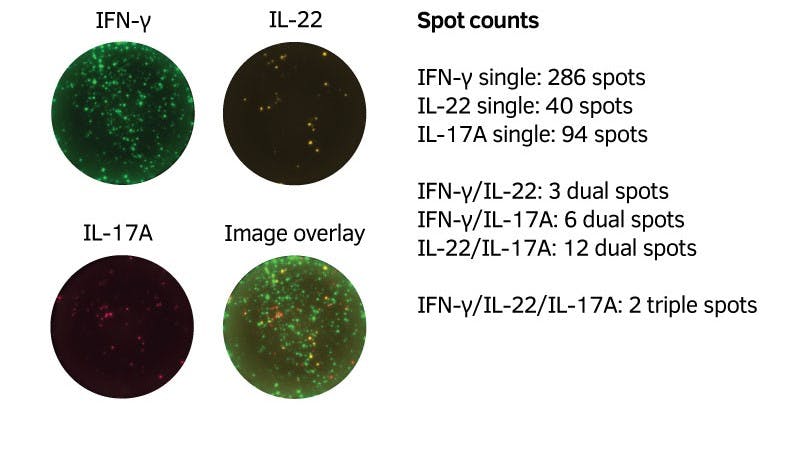
Mouse splenocytes (100,000 cells/well) were incubated for 48 hours in the absence or presence of ConA. The number of cells secreting IFN-γ, IL-22 and IL-17A was analyzed by FluoroSpot. Single cytokine secreting cells as well as double- and triple-secreting cells were determined. Spot center was used to identify spots from double- and triple-secreting cells, visualized in an image overlay of LED490, FITC (IFN-γ) and LED550, Cy3 (IL-22) and LED640, Cy5 (IL-17A). Spot analysis was made with Mabtech IRIS.


IFN-γ
| Analyte description | Interferon-γ (IFN-γ) is the only type II interferon. This proinflammatory cytokine is secreted by activated T cells and NK cells. It activates macrophages and endothelial cells and regulates immune responses by affecting APCs, T cells, and B cells. Production of IFN-γ by helper T cells and cytotoxic T cells is a hallmark of the Th1-type phenotype. Thus, high-level production of IFN-γ is typically associated with effective host defense against intracellular pathogens. |
| Alternative names | Interferon-γ, Interferon-gamma, IFN-γ, IFN-gamma, IFN-g, IFNg, If2f, Ifg |
| Cell type | T cell, Tc, Th1, NK cell |
| Gene ID | 15978 |
IL-22
| Analyte description | The cytokine interleukin 22 (IL-22) is mainly produced by activated CD4 T helper 17 (Th17) and T helper 22 (Th22) cells. The IL-22 receptor is expressed on non-immune cells, particularly epithelial cells and keratinocytes, and IL-22 promotes innate immune responses versus bacterial infections in these target cells. In addition, Th22 cells have been shown to play a role in the pathophysiology of several human skin diseases. |
| Alternative names | Interleukin-22, IL-22, IL22, IL-22a, ILTIFa, If2b1, Iltif |
| Cell type | T cell, Th1, Th17, Tfh |
| Gene ID | 50929 |
IL-17A
| Analyte description | Interleukin 17A (IL-17A) is a potent proinflammatory cytokine produced by activated Th17 (T helper 17) cells and certain cells belonging to the innate immune system. In mice, IL-17 has also been shown to be produced by activated CD8 T cells and γδ T cells. Th17 cells play an important role in autoimmune diseases and protection against bacteria and fungi. IL-17A acts on a broad range of cell types to induce the expression of cytokines, chemokines, and metalloproteinases. As a result, secretion of IL-17A promotes inflammatory responses, which leads to the recruitment of neutrophils, enhancement of antibody production, and activation of T cells. Increased expression of IL-17A is seen in autoimmune diseases such as multiple sclerosis and rheumatoid arthritis. It is also associated with asthma, psoriasis, cancer, and transplant rejection. |
| Alternative names | Interleukin 17A, IL-17A, IL17A, Ctla-8, Ctla8, IL-17, Il17 |
| Cell type | Th17 |
| Gene ID | 16171 |