FluoroSpot Plus: Mouse IFN‑γ/IL‑2

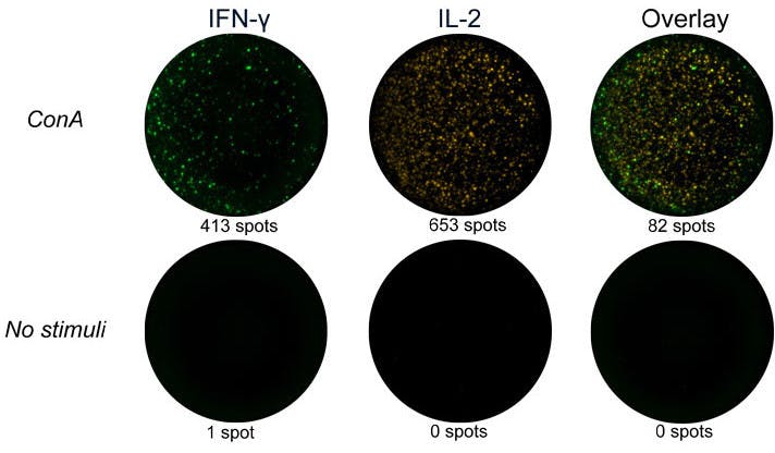
FluoroSpot Plus: Mouse IFN‑γ/IL‑2
Components
| Plate | Pre-coated FluoroSpot plate (mAbs AN18 and 1A12) |
| Detection mAbs | Anti-IFN-γ mAb (MTR7), BAM |
| Anti-IL-2 mAb (5H4), biotin | |
| Fluorophore conjugates | Anti-BAM mAb, 490 |
| SA-550 | |
| Buffer/Solution | FluoroSpot enhancer |
| Co-stimulus | Anti-CD28 mAb |
In stock
Delivery 4-9 business days
Shipping $0


IFN-γ
| Analyte description | Interferon-γ (IFN-γ) is the only type II interferon. This proinflammatory cytokine is secreted by activated T cells and NK cells. It activates macrophages and endothelial cells and regulates immune responses by affecting APCs, T cells, and B cells. Production of IFN-γ by helper T cells and cytotoxic T cells is a hallmark of the Th1-type phenotype. Thus, high-level production of IFN-γ is typically associated with effective host defense against intracellular pathogens. |
| Alternative names | Interferon-γ, Interferon-gamma, IFN-γ, IFN-gamma, IFN-g, IFNg, If2f, Ifg |
| Cell type | T cell, Tc, Th1, NK cell |
| Gene ID | 15978 |
IL-2
| Analyte description | Interleukin 2 (IL-2) is predominantly produced by activated T cells. IL-2 promotes the proliferation, differentiation, and survival of antigen-activated T cells and NK cells. After antigen stimulation, the cytokine also promotes the differentiation of T cells into effector and memory T cells. Therefore IL-2 plays an important role in the body's response to infection. IL-2 is also key to tolerance, as it promotes the differentiation of certain immature T cells into regulatory T cells in the thymus, ultimately preventing autoimmune diseases. |
| Alternative names | Interleukin-2, Interleukin 2, IL-2, IL2, T cell growth factor, Il-2 |
| Cell type | T cell, Th1, Th2, Th17, Tfh |
| Gene ID | 16183 |