FluoroSpot Plus: Mouse IFN‑γ/IL‑17A

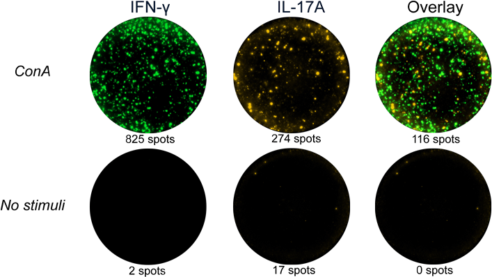
FluoroSpot Plus: Mouse IFN‑γ/IL‑17A
Components
| Plate | Pre-coated FluoroSpot plate (mAbs AN18 and IL17-I) |
| Detection mAbs | Anti-IFN-γ mAb (MTR7), BAM |
| Anti-IL-17A mAb (MT2270), biotin | |
| Fluorophore conjugates | Anti-BAM mAb, 490 |
| SA-550 | |
| Buffer/Solution | FluoroSpot enhancer |
| Co-stimulus | Anti-CD28 mAb |
Low stock
Delivery within 3-4 weeks
Shipping $0


IFN-γ
| Analyte description | Interferon-γ (IFN-γ) is the only type II interferon. This proinflammatory cytokine is secreted by activated T cells and NK cells. It activates macrophages and endothelial cells and regulates immune responses by affecting APCs, T cells, and B cells. Production of IFN-γ by helper T cells and cytotoxic T cells is a hallmark of the Th1-type phenotype. Thus, high-level production of IFN-γ is typically associated with effective host defense against intracellular pathogens. |
| Alternative names | Interferon-γ, Interferon-gamma, IFN-γ, IFN-gamma, IFN-g, IFNg, If2f, Ifg |
| Cell type | T cell, Tc, Th1, NK cell |
| Gene ID | 15978 |
IL-17A
| Analyte description | Interleukin 17A (IL-17A) is a potent proinflammatory cytokine produced by activated Th17 (T helper 17) cells and certain cells belonging to the innate immune system. In mice, IL-17 has also been shown to be produced by activated CD8 T cells and γδ T cells. Th17 cells play an important role in autoimmune diseases and protection against bacteria and fungi. IL-17A acts on a broad range of cell types to induce the expression of cytokines, chemokines, and metalloproteinases. As a result, secretion of IL-17A promotes inflammatory responses, which leads to the recruitment of neutrophils, enhancement of antibody production, and activation of T cells. Increased expression of IL-17A is seen in autoimmune diseases such as multiple sclerosis and rheumatoid arthritis. It is also associated with asthma, psoriasis, cancer, and transplant rejection. |
| Alternative names | Interleukin 17A, IL-17A, IL17A, Ctla-8, Ctla8, IL-17, Il17 |
| Cell type | Th17 |
| Gene ID | 16171 |