Dear Mabtech staff and researchers,
I am conducting MHV(murine hepatitis virus) Surface protein peptide-specific T cell ELISPOT assay.
using Elispot plate: M8IPS4510 | MultiScreenHTS IP Filter Plate, 0.45 µm, clear, non-sterile
Primary IFN-g antibody (capture): BD cat no. 554430, final concentration in Elispot plate is 3.0 µg/ml
Secondary IFN-g antibody (biotinylated; for detection): BD cat no. 554410, final concentration in Elispot plate is 2.0 µg/ml
Positive control: PHA-M (SIGMA L8902-5MG), final concentration in Elispot plate: 2 µg/ml
I seeded 1,000,000 cells/well splenocytes from MHV infected mice.
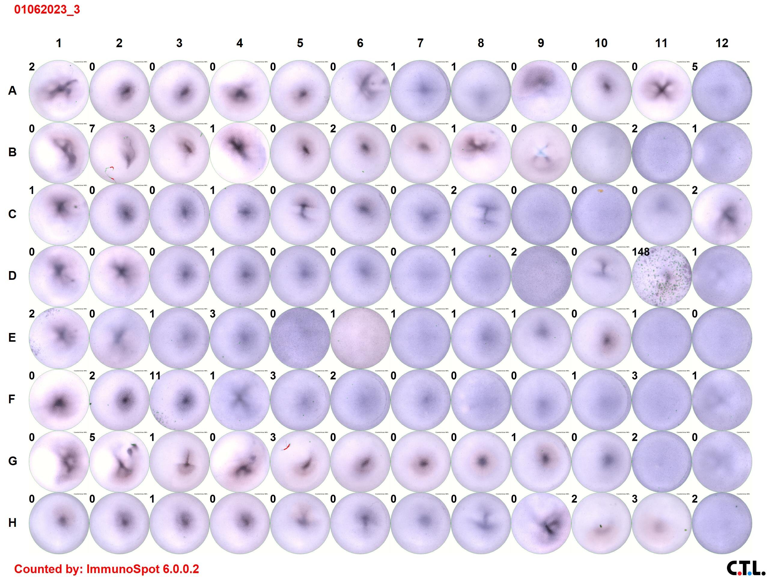
Attached is the image of the result.
As we can see the images, the center of some wells have purple area. Have you ever had such experiences?
I thought it was because of ethanol when pre coating. Do you think is it meaningful without ethanol ELISPOT?
Finally, we could not see many spot in positive control, PHA (H11 and H12 in the image), but other some wells have many spots. Do you know that PHA is correct as mice splenocytes?
Thank you,
Jun