Hi, dear support team of MABTECH
Our team are working on an Elispot IFN-g with chicken splenocytes using the MABTECH kit ELISpot PLUS: Chicken IFN-g (ALP). We have been reading this forum in order to learn from your advice. Now we have some specific doubts that we would like to clarify.
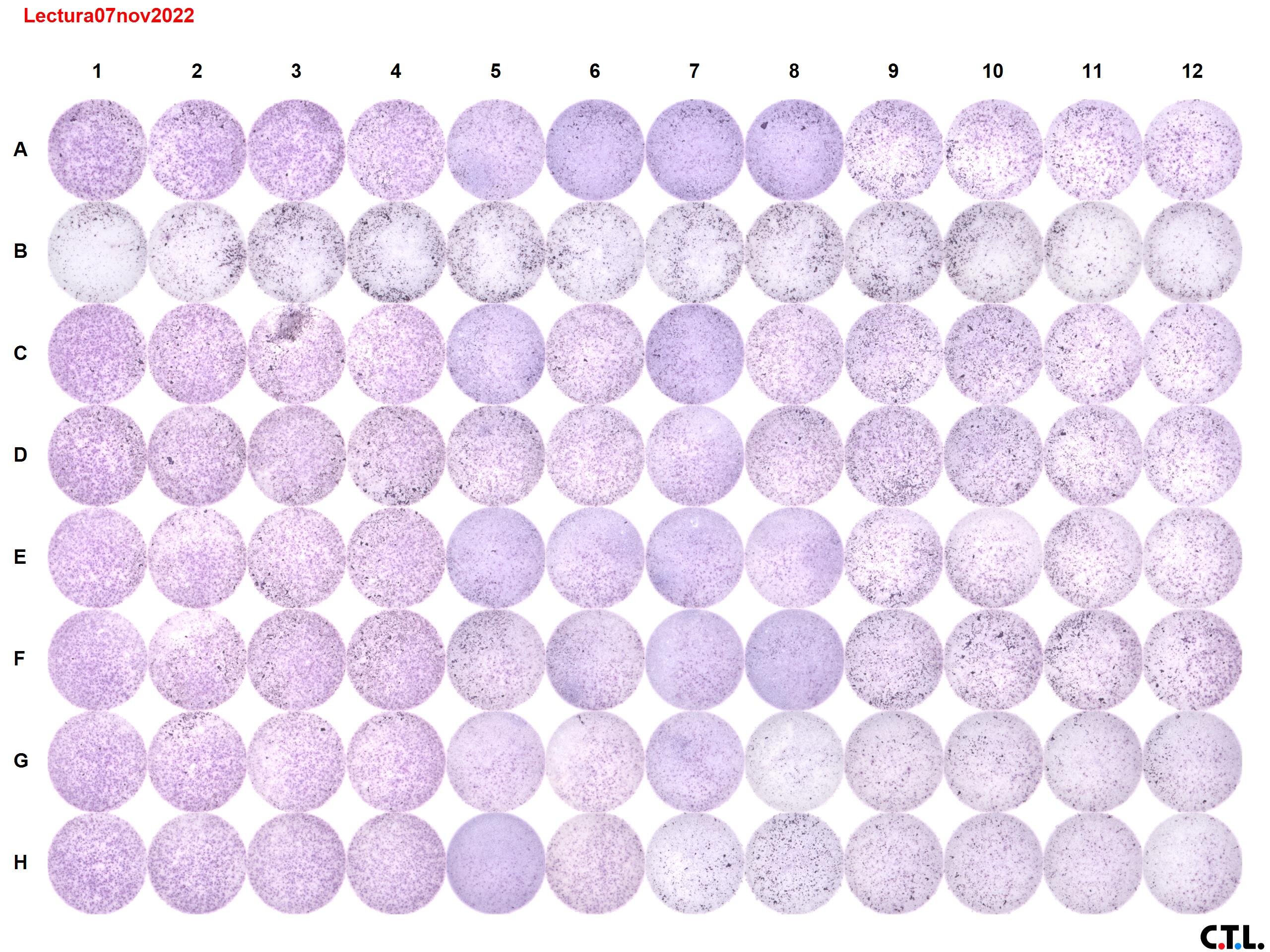
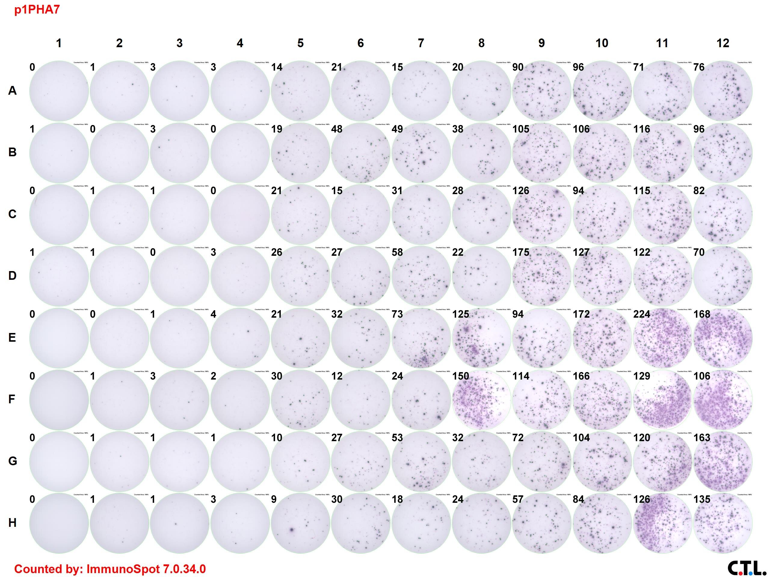
-We are following the exact instructions of the kit and in the past experiments, we had really nice results for the standardization of the conditions (Standarizationp1PHA7.jpg). In our last experiment (Lectura07no2022.jpg), we saw two things that we hadn't seen before. In almost all our wells we have kind of a precipitate. It can be seen clearly in row B, where we have no stimuli to the cells. Additionally, in some of our groups, we have membrane staining in some wells of columns 6-8. Could you give us any advice or do you have any idea of what could be happening?
-Related to the spot number per well: Do you recommend a specific range of spot number per well? We have prove different cell concentrations (from 100,000-500,000 cells/well) and we selected 300,000 cells/well because we had <500 spots. Recently, we prove perfusion as a new method to set apart the splenocytes. We got ~99% of viability, so now we have much more spots than initially with cell grinder (80-90% viability).
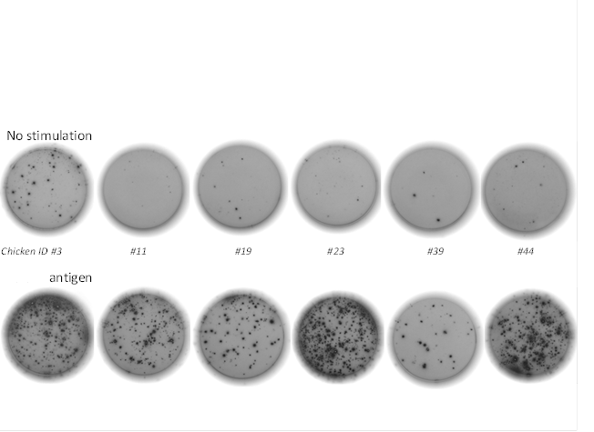
-Finally, we would like to ask about the negative controls. We had been using a synthetic peptide with a random sequence that in appearance is not related to our antigen (in fact it have 0 % identity in sequence) but at the ELISPOT assay we are detecting that it produce almost the same reaction than our synthetic peptides related to our antigens (100 % identity to the antigen). Do you have any comment for these or could you give us a recommendation for negative control?
Thank you in advance!
Regards