PepPool: CMV (CD4 and CD8), human

PepPool: CMV (CD4 and CD8), human

3619-1

Publications: 8
Documents
Questions?
Validated for our assays.

Content

Peptide pool with 42 peptides, 25 ug of each peptide. Supplied in one vial (lyophilized).

In stock

Delivery 4-9 business days

Shipping $0

Product specifications

Intended use

This peptide pool is intended for studying immune responses against CMV in immunoassays such as ELISpot, FluoroSpot, and flow cytometry. It induces cytokine secretion from antigen-specific human CD4 and CD8 T cells. For research use only. Not for use in diagnostic procedures.

Serum/Plasma samples

Description
The CMV peptide pool induces secretion of e.g., IFN-γ, IL-2, IL-5, and IL-13 from human CD4 and CD8 T cells. It contains 42 peptides from cytomegalovirus. The antigens represented in the peptide pool are pp50, pp65, IE1, IE2, and envelope glycoprotein B. 28 of the peptides are MHC class I-restricted and 14 are MHC class II-restricted. The synthetic peptides are >95% pure.

HLA types covered by the CMV pool:
HLA class I: HLA-A1, HLA-A2, HLA-A3, HLA-A11, HLA-A23, HLA-A24, HLA-A26, HLA-A30, HLA-B7, HLA-B8, HLA-B18, HLA-B27, HLA-B35, HLA-B40/60, HLA-B41, HLA-B44, HLA-B57/58, HLA-C7. HLA class II: HLA-DRB1*01, 03, 04, 05, 07, 08, 11, 15, 20, 24, 53, HLA-DPw2, HLA-DQB, HLA-DP3*14, 20

Recommendation

Product details

ProductPepPool: CMV (CD4 and CD8), human
ApplicationT cell stimulation, ELISpot, FluoroSpot, Flow cytometry
ReactivityHuman
Supplied in

Lyophilized

Contents

Peptide pool with 42 peptides, 25 ug of each peptide. Supplied in one vial (lyophilized).

Shipping and Storage

Shipping

Shipped at ambient temperature.

Storage

Store at -20 °C or below upon receipt.

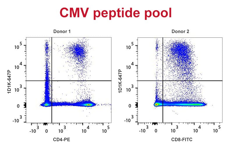
Human PBMC stimulated with CMV peptide pool (2 ug/ml) and anti-CD28 antibody (0.1 ug/ml) for 16 hours (both available from Mabtech) in the presence of Brefeldin A. In donor 1, CD4+ cells start expressing IFN-γ following CMV peptide pool stimulation whereas donor 2 responds by production of IFN-γ by CD8+ cells. Cells were stained with CD4-PE and CD8-FITC prior to fixation and permeabilization using 4% paraformaldehyde and saponin. IFN-γ was detected using 1D1K-PF647P (Mabtech). The dot plots derive from gated events of typical lymphocyte characteristics in forward and side light scatter. Flow cytometry was performed on a BD FACSVerse system.

Loading publications...
1 / 1